Collagen Corneal Cross Linking
"Busy day at work today Breanna?"
"Sure was, we had a cross linking procedure so that made the day fly by."
"Ah okay... and what on earth is a cross linking??" *Confused look on face*
This is a conversation that I've struggled to answer simplistically many times when I catch up with friends or family after work over the dinner table...
So this blog post is all about 'Collagen Corneal Cross Linking' also known as 'Cross Linking' or 'CXL'
 I thought it was perfect timing to share my experiences with cross linking because from May 1st this year the procedure will be approved on the Medicare PBS. This is super exciting for us in the world of Ophthalmology as it means patients will be eligible for a rebate from this minor operation. This highlights that there have now been a sufficient number of clinical trials completed to prove it's success. I have actually even been involved in auditing the efficacy of CXL at my workplace and it's been incredible to examine the benefits to patients.
I'm really excited about this post as I want to share with you just how rewarding and stimulating it is as an Orthoptist to be apart of a treatment that aims to halt progression of a corneal disease and prevent the need for a corneal transplant.
So let's unravel what 'CXL' really means!
First I think to understand the treatment of CXL you need to have insight into what eye condition this procedure is used as a management for. The eye condition responsible is called 'Keratoconus' - bit of a tounge twister so we will call it KC!
KC effects the cornea which is the clear window on the front of the eye. The normal cornea is round alike a basketball where as a patient with KC will have an abnormally shaped cornea similar to a football.
So where does CXL fit into all of this? CXL is a procedure designed to HALT the progression of KC before it becomes too advanced and aim to prevent the need for a graft. It is much less invasive than a transplant and the recovery time is much quicker.
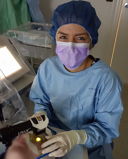
So what role does an Orthoptist play in CXL? At the private clinic I work at we actually have two Orthoptists assisting during a cross linking, one Orthoptist will be fully sterile and will set up the treatment room by adjusting the microscope and UV light as well as preparing all the ophthalmic surgical instruments.
 |
| Ophthalmic surgical instruments used in CXL |
The Ophthalmologist does the initial part of the treatment which involves removing the surface layer of the cornea (epithelium) and the scrubbed in Orthoptist will pass the surgeon ophthalmic instruments in a particular order to complete this process.
After the Ophthalmologist has removed the surface cells, a yellow eye drop called Riboflavin is administered onto the eye. Riboflavin is a Vitamin B2 (fun fact: it's actually found in Vegemite) and it's used to help strengthen the collagen bonds in the cornea and ultimately halt progression of KC. So every two minutes for half an hour an Orthoptist will instil the Riboflavin drops and also periodically take ultrasound measurements of the thickness of the cornea to ensure it stays within a certain parameter.
Once the first 30 minutes are over the Ophthalmologist will check that the Riboflavin has soaked into the eye properly and then the second part of the treatment begins. This involves an Ultraviolet light positioned over the eye to activate the Riboflavin. The Orthoptist will again administer Riboflavin eye drops every 2 minutes and also complete ultrasound measurements while ensuring the UV light is focused appropriately over the eye. The second half of the treatment can be more challenging because if the patients corneal ultrasounds become too thin then swelling of the cornea with water is required and sometimes even a contact lens is placed on the eye to protect the posterior part of the cornea.
At the end of the treatment the Orthoptist will put in a series of healing drops and also a bandage contact lens which stays in the patients eye for a week to allow the surface cells to regenerate. We usually complete one day, one week, one month and three month post-operative appointments to monitor the recovery. At the three month mark we can usually have a good indication if the patient has responded well to the cross linking.
VWA-LAH. Thats cross linking in a nutshell!
I think I have found it most rewarding to be able to guide patients amicably through a cross linking, as you can imagine it would be very daunting having cells removed from the front of you eye while you are completely awake. Most of these patients are quite young and can be very sensitive when it comes to their eyes (understandably). I enjoy being able to help patients overcome any anxieties by providing support and education about their corneal condition. Most of all for me it's very fulfilling to review patients after a few months and assess their eyes to find no worsening of the condition and to know you have been apart of this process.
Eye'm out,
B


Hi B, I loved learning about KC. Can you let us know what the average age a person is diagnosed with KC?
ReplyDeleteI can’t wait for your next post!
Thanks, RB.
Hey Cynthia!
DeleteThank you for your comment! KC usually begins during puberty (as a teenager) and stabilises around the age of 25 unless the patient has had poorly fitted contact lenses or excessive rubbing of the eyes due to allergy related symptoms. Please let me know if you have any other questions or have any topics you would like me to blog about.
Eye'm out,
B